Echocardiography (ECHO)

Overview
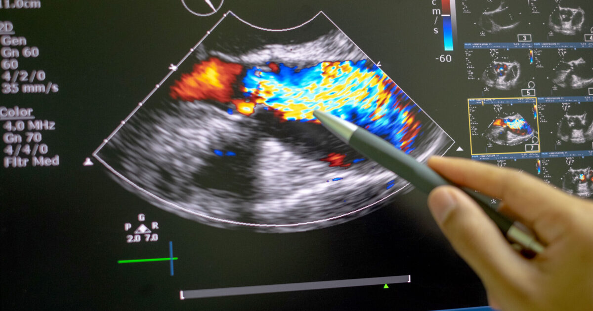
This test uses 1.5 to 7.5 MHz sound waves to examine the heart and produce a 2D or 3D image of the heart’s internal structure called the echocardiogram.

An echocardiogram, commonly known as ‘echo’ provides information about the heart’s size, the size and functioning of the heart’s valve, the size of the heart’s chambers, the thickness of cardiac walls, the pumping activity of the heart, growth of any tumor in the heart, and the blood flow pattern. It is referred by cardiologists if there are any abnormalities in the Electrocardiogram (ECG/EKG), shortness of breath, chest pain (angina), or any other underlying medical conditions indicating a cardiac problem.

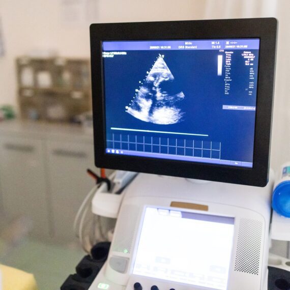
The echocardiography machine contains a transducer or probe that contains piezoelectric crystals for interchanging electric impulse into ultrasound and vice-versa, a central processing unit with a control panel for receiving signals from the transducer and processing and analyzing the signal, a monitor to display an image of the heart and printer for printing the produced electrocardiogram.
Echocardiography

What We Do?

At Nayab Labs & Diagnostic Centre, we offer comprehensive echocardiogram (echo) services to assess and monitor heart health. Our advanced echo tests use ultrasound technology to provide real-time images of the heart’s structure and function, assisting doctors in diagnosing conditions such as heart valve issues, abnormalities in heart muscle function, and irregular blood flow patterns.

With our state-of-the-art equipment and skilled technicians, we ensure that each echo test is performed with precision, providing reliable insights into cardiac health for effective medical evaluation and treatment planning.
echocardic
Echocardiographo

Types of Echocardiography (Echo)


Based on the approach followed to develop an echocardiogram and/or the types of information needed, echocardiography can be classified into several types. Some of them are listed below:

1. Transthoracic Echocardiography (TTE)
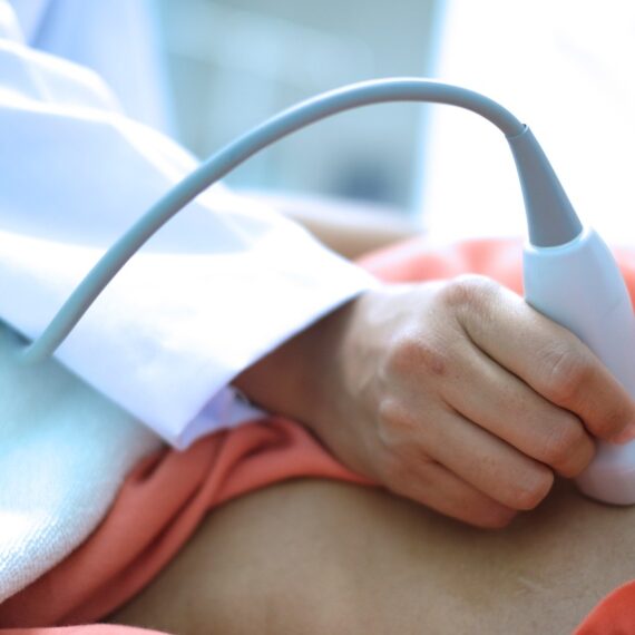
It is the standard echocardiography in which the transducer is placed on the chest wall.

2. Transesophageal Echocardiography
It is an echocardiography technique in which a specialized transducer is passed through the mouth to the esophagus, and a scan is done by passing the sound wave internally.

3. Stress Echocardiography
It is the type of echo that combines the ultrasound scanning of the heart before and after exercise or stress to study the heart’s structure and functioning during physical stress. In this test, echo is done before exercise and after about 10 to 15 minutes of exercise.

4. Intracardiac Echocardiography
It is an invasive type of echocardiography in which a catheter with an ultrasound transducer at its tip is inserted in the femoral vein and passed up to the heart to produce an ultrasound-scanning image of the heart.

Procedure of Transthoracic Echocardiography (Echo)
The patient is asked to lie flat on a bed (examination table) with the chest exposed. (Any metallic objects, jewelry, and electronic devices are asked to be removed.)
The technician will apply the gel on the chest so that the sound waves can easily pass to the heart.
The echo probe or transducer is placed at a specific location on the chest, and ultrasonic sound waves are passed through the transducer. The transducer will also receive the reflected sound (echo) and will send it to the machine for processing.
Once the desired echocardiogram is obtained, the machine is turned off, the transducer is removed, and the gel is wiped. The echocardiogram is printed and submitted to the cardiologist for interpretation.


chemlabs-38

Preparing for Test Help Ensure It Go Smoothly.

To reduce your wait time, complete the personal information on the form in advance.